
Figure 1 – Cross section of a pixel on a color digital camera CCD sensor. From the Wikicommons and in the public domain.
We have been talking about camera lenses, those big objects in the front of your camera that form the image. But remember that, as we discussed when we considered CCD arrays, that there is a second type of lens in your imaging system. These are the microlens array (see Figure 1 ) that lie atop the CCD elements. The purpose of these microlenses, you may recall, is not specifically to image but rather to collect as much light as possible and direct it to the photosensitive array elements. On camera sensor chips there is often considerable dead space and the task of these lens is to collect all this light and bring it to the sensor. This becomes really essential as you put more and more pixels onto the chip, and you need to maximize the signal because of the limited surface area and well-depth.
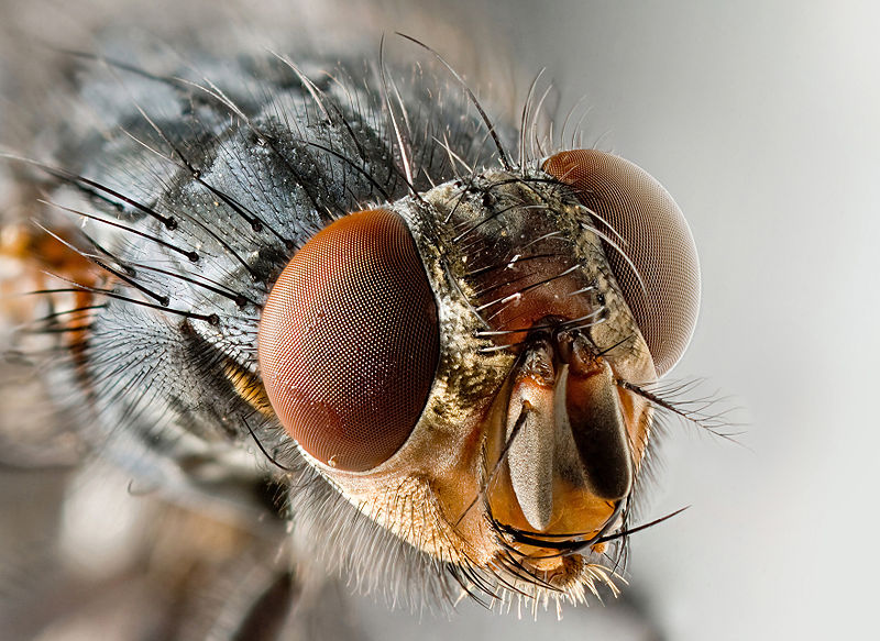 This concept of a sensor element or pixel which reads a single point and outputs a grey level, or color, but not an image, is, of course a copy of compound eyes in nature. Figure 2 shows a the compound eye of a fly. Figure 3 shows the microlenslet array of the house fly’s eye in a scanning electron microscope. The compound eye of arthropods provides tremendous field of view and makes the animal very sensitive to motion, as anyone who has tried to swat a fly or mosquito soon realizes.
The situation with the compound insect eye is not quite identical to that of the microlens in a CCD array. Insects eyes are closer to what would happen if each microlenslet covered several pixels; so that you could form a crude image. But if you think of that situation light would be coming in from everywhere and each pixel array, called an ommatidium, having a low resolution image of the whole scene. This is the popular view of movies etc., but not really what happens. The ommatidia are designed to limit the amount of light coming in from extremen angles. As a result the image is only of the scene immediately perpendiculr to ommatidium. So what the insect sees is a tiled view of its world. Part of the key to all of this is that something that moves between ommatidia is rapidly detected, which is what the insect needs to find food and escape being eaten.
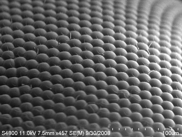
Figure 3 – Scanning electron micrograph of a house fly’s eye showing the microlens array. From the Wikimedia Commons by United Nation and in the public domain under creative commons license.
The resolution of such eyes is however, very limited. For instance, the resolving power of the honey bee’s eye is only 1/60th that of the human or vertebrate eye. What a vertebrate can resolve at 60 feet (18 m) the bee can only resolve at a distance of one foot (0.3 m). To see with a resolution comparable to our vertebrate eyes, humans would require compound eyes about 22 m or sixty 70 feet in diameter. All of this brings to mind Vincent Price as “The Fly, 1958.” “Phillipe help me!” Although I have to say that I prefer the 1986 remake with Jeff Goldblum and Geena Davis.